Holographic microscopy

We use a fast imaging method called holographic microscopy to watch self-assembling systems. In a holographic microscope, the sample is illuminated by laser light, and the resulting image (or hologram) can be used to determine the 3D structure, position, and orientation of a microscopic sample. We build new holographic microscopes and develop software to analyze the holograms.

An introduction to holography

Dennis Gabor developed holography in the 1940s as a way to improve the resolving power of electron microscopes. However, his ideas apply to imaging with any coherent source. With the development of lasers in the 1960s, it became possible to do holographic recording and reconstruction with visible light and photographic film. Later, when CCD and CMOS cameras arrived on the scene, holograms could be recorded digitally. Today, with high-speed computers, we can not only record holograms digitally, but also reconstruct them digitally as well.

To understand holography, we need to consider the wave nature of light. At any point in space, a light wave can be described by an amplitude and phase. Whereas a conventional photograph captures the intensity of light reflected or scattered from an object, a hologram can capture both amplitude and phase. This is because the hologram is recorded by using two light beams, one of which (called the object beam) is scattered from the object, and other (called the reference beam) is aimed at the camera. If the two waves are coherent (meaning the phase is well-defined at each point in space), they can interfere, or beat, with each other to produce interference fringes, like this:
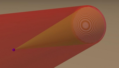
Light scatters off a particle, and an interference pattern i.e. hologram is created.

In the picture above, the small blue particle is illuminated by the red beam. It scatters light into an object beam, shown as a cone radiating from the sphere. That object beam interferes with the red beam to produce a fring pattern, shown on the right side of the image.

The fringes encode information about the phase of light and, implicitly, about the position and structure of the object. That information can be recovered through reconstruction. The simplest way to recover the 3D information is to record the hologram as an intensity pattern on a camera. If, say, we take the image and print it on photographic film, we can then shine light back through it. The hologram will diffract the light so that an image of the object will become visible:

 A reference beam propagating back through a hologram will be focussed to a point close to where the original object was

This can most easily be understood if one imagines recording the hologram of a spherically scattered wave (like the light scattered from a microscopic particle). If one interferes that spherical wave coming from the object with a plane wave, a pattern of concentric rings will be observed. These fringes will resemble a Fresnel zone plate. And, just like a Fresnel zone plate, the fringes will focus a plane wave illuminating it to a point.

In our lab, we build holographic microscopes, and we develop techniques for doing 3D reconstruction on a computer.  Below are some of the techniques we use.

In-line holographic microscopy

The in-line holographic microscope operates as described above. A laser illuminates the sample, which is typically a colloidal or biological sample suspended in a liquid.  Some of that light passes through the sample and acts as the reference beam, and some light is scattered by the object. We record the hologram on a CMOS camera, and we reconstruct it on a computer to recover 3D information about the sample.  We can do the numerical equivalent of shining the reference beam back on the hologram and looking at the diffracted image, or, if we know what our object is beforehand, we can numerically fit a scattering model to the observed hologram using our software package, HoloPy.

We are currently able to track micron-scale spheres to a precision of less than 1 nm precision in all three dimensions, at  high frame rates (over 5000 frames/second). We are also able to track clusters of spheres, and non-spherical particles and their rotations in 3-dimensions to a spatial precision of about 10 nm . Below is a video showing (on the left) a series of holograms taken of a 0.37 x 1.05 um polystyrene ellipsoid diffusing in water and (on the right) a rendering of the particle, showing the 3D position and orientation we infer from fitting a model to the recorded holograms. The movie is sped up by a factor of 5 from real time.

Publications

Wang, A. ; McGorty, R. ; Kaz, D. M. ; Manoharan, V. N. Contact-line pinning controls how quickly colloidal particles equilibrate with liquid interfaces. Soft Matter 2016, 12, 8958-8967. Publisher's VersionAbstract
Previous experiments have shown that spherical colloidal particles relax to equilibrium slowly after they adsorb to a liquid-liquid interface, despite the large interfacial energy gradient driving the adsorption. The slow relaxation has been explained in terms of transient pinning and depinning of the contact line on the surface of the particles. However, the nature of the pinning sites has not been investigated in detail. We use digital holographic microscopy to track a variety of colloidal spheres---inorganic and organic, charge-stabilized and sterically stabilized, aqueous and non-aqueous---as they breach liquid interfaces. We find that nearly all of these particles relax logarithmically in time over timescales much larger than those expected from viscous dissipation alone. By comparing our results to theoretical models of the pinning dynamics, we infer the area per defect to be on the order of a few square nanometers for each of the colloids we examine, whereas the energy per defect can vary from a few $kT$ for non-aqueous and inorganic spheres to tens of $kT$ for aqueous polymer particles. The results suggest that the likely pinning sites are topographical features inherent to colloidal particles---surface roughness in the case of silica particles and grafted polymer ``hairs'' in the case of polymer particles. We conclude that the slow relaxation must be taken into account in experiments and applications, such as Pickering emulsions, that involve colloids attaching to interfaces. The effect is particularly important for aqueous polymer particles, which pin the contact line strongly.
Rahmani, A. M. ; Wang, A. ; Manoharan, V. N. ; Colosqui, C. E. Colloidal particle adsorption at liquid interfaces: capillary driven dynamics and thermally activated kinetics. Soft Matter 2016, 12, 6365-6372. Publisher's VersionAbstract
The adsorption of single colloidal microparticles (0.5–1 μm radius) at a water–oil interface has been recently studied experimentally using digital holographic microscopy [Kaz et al., Nat. Mater., 2012, 11, 138–142]. An initially fast adsorption dynamics driven by capillary forces is followed by an unexpectedly slow relaxation to equilibrium that is logarithmic in time and can span hours or days. The slow relaxation kinetics has been attributed to the presence of surface “defects” with nanoscale dimensions (1–5 nm) that induce multiple metastable configurations of the contact line perimeter. A kinetic model considering thermally activated transitions between such metastable configurations has been proposed [Colosqui et al., Phys. Rev. Lett., 2013, 111, 028302] to predict both the relaxation rate and the crossover point to the slow logarithmic regime. However, the adsorption dynamics observed experimentally before the crossover point has remained unstudied. In this work, we propose a Langevin model that is able to describe the entire adsorption process of single colloidal particles by considering metastable states produced by surface defects and thermal motion of the particle and liquid interface. Invoking the fluctuation dissipation theorem, we introduce a drag term that considers significant dissipative forces induced by thermal fluctuations of the liquid interface. Langevin dynamics simulations based on the proposed adsorption model yield close agreement with experimental observations for different microparticles, capturing the crossover from (fast) capillary driven dynamics to (slow) thermally activated kinetics.
Dimiduk, T. G. ; Manoharan, V. N. Bayesian approach to analyzing holograms of colloidal particles. Optics Express 2016, 24, 24045–24060. Publisher's VersionAbstract

We demonstrate a Bayesian approach to tracking and characterizing colloidal particles from in-line digital holograms. We model the formation of the hologram using Lorenz-Mie theory. We then use a tempered Markov-chain Monte Carlo method to sample the posterior probability distributions of the model parameters: particle position, size, and refractive index. Compared to least-squares fitting, our approach allows us to more easily incorporate prior information about the parameters and to obtain more accurate uncertainties, which are critical for both particle tracking and characterization experiments. Our approach also eliminates the need to supply accurate initial guesses for the parameters, so it requires little tuning.

Wang, A. ; Garmann, R. F. ; Manoharan, V. N. Tracking E. coli runs and tumbles with scattering solutions and digital holographic microscopy. Optics Express 2016, 24, 23719–23725. Publisher's VersionAbstract

We use in-line digital holographic microscopy to image freely swimming E. coli. We show that fitting a light scattering model to E. coli holograms can yield quantitative information about the bacterium&\#x02019;s body rotation and tumbles, offering a precise way to track fine details of bacterial motility. We are able to extract the cell&\#x02019;s three-dimensional (3D) position and orientation and recover behavior such as body angle rotation during runs, tumbles, and pole reversal. Our technique is label-free and capable of frame rates limited only by the camera.

Goldfain, A. M. ; Garmann, R. F. ; Jin, Y. ; Lahini, Y. ; Manoharan, V. N. Dynamic Measurements of the Position, Orientation, and DNA Content of Individual Unlabeled Bacteriophages. The Journal of Physical Chemistry B 2016, 120, 6130–6138. Publisher's VersionAbstract

A complete understanding of the cellular pathways involved in viral infections will ultimately require a diverse arsenal of experimental techniques, including methods for tracking individual viruses and their interactions with the host. Here we demonstrate the use of holographic microscopy to track the position, orientation, and DNA content of unlabeled bacteriophages (phages) in solution near a planar, functionalized glass surface. We simultaneously track over 100 individual λ phages at a rate of 100 Hz across a 33 μm × 33 μm portion of the surface. The technique determines the in-plane motion of the phage to nanometer precision, and the height of the phage above the surface to 100 nm precision. Additionally, we track the DNA content of individual phages as they eject their genome following the addition of detergent-solubilized LamB receptor. The technique determines the fraction of DNA remaining in the phage to within 10% of the total 48.5 kilobase pairs. Analysis of the data reveals that under certain conditions, λ phages move along the surface with their heads down and intermittently stick to the surface by their tails, causing them to stand up. Furthermore, we find that in buffer containing high concentrations of both monovalent and divalent salts, λ phages eject their entire DNA in about 7 s. Taken together, these measurements highlight the potential of holographic microscopy to resolve the fast kinetics of the early stages of phage infection.

Perry, R. W. Internal Dynamics of Equilibrium Colloidal Clusters, 2015. Publisher's VersionAbstract

Colloidal clusters, aggregates of a few micrometer-sized spherical particles, are a model experimental system for understanding the physics of self-assembly and processes such as nucleation. Colloidal clusters are well suited for studies on these topics because they are the simplest colloidal system with internal degrees of freedom. Clusters made from particles that weakly attract one another continually rearrange between different structures. By characterizing these internal dynamics and the structures connected by the rearrangement pathways, we seek to understand the statistical physics underlying self-assembly and equilibration.

In this thesis, we examine the rearrangement dynamics of colloidal clusters and analyze the equilibrium distributions of ground and excited states. We prepare clusters of up to ten microspheres bound by short-range depletion interactions that are tuned to allow equilibration between multiple isostatic arrangements. To study these clusters, we use bright-field and digital holographic microscopy paired with computational post-processing to amass ensemble-averaged and time-averaged probabilities.

We study both two-dimensional (2D) and three-dimensional (3D) clusters composed of either one or two species of particles. To learn about geometrical nucleation barriers, we track rearrangements of particles within freely rotating and translating 3D clusters. We show that rearrangements occur on a timescale of seconds, consistent with diffusion-dominated internal dynamics. To better understand excited states and transition pathways, we track hundreds of rearrangements between degenerate ground states in 2D clusters. We show that the rearrangement rates can be understood using a model with two parameters, which account for the diffusion coefficient along the excited-state rearrangement pathways and the interaction potential. To explore new methods to control self-assembly, we analyze clusters of two species with different masses and different interactions. We find that the interactions allow for control over the intracluster placement of each species, while the masses have no influence. To provide a theoretical framework for understanding these observations, we derive the classical partition function of colloidal clusters in terms of translational, rotational, and vibrational degrees of freedom. We show that the masses of the particles enter the partition function through the kinetic energy but have no effect on the probabilities of states that differ only in where the masses are placed. This result is consistent with our experiments.

Overall, this work shows that the equilibrium distribution of self-assembled colloidal clusters is well-modeled by classical statistical physics, and that the rearrangement dynamics of colloidal clusters can be understood by incorporating diffusion and the effect of the interaction potential. Because both the structures and dynamics can be accurately predicted, these clusters are a promising system for self-assembling novel materials and for studying the emergence of phase transitions.

Dimiduk, T. G. ; Perry, R. W. ; Fung, J. ; Manoharan, V. N. Random-subset fitting of digital holograms for fast three-dimensional particle tracking [Invited]. Applied Optics 2014, 53, G177-G183. Publisher's VersionAbstract

Fitting scattering solutions to time series of digital holograms is a precise way to measure three-dimensional dynamics of microscale objects such as colloidal particles. However, this inverse-problem approach is computationally expensive. We show that the computational time can be reduced by an order of magnitude or more by fitting to a random subset of the pixels in a hologram. We demonstrate our algorithm on experimentally measured holograms of micrometer-scale colloidal particles, and we show that 20-fold increases in speed, relative to fitting full frames, can be attained while introducing errors in the particle positions of 10 nm or less. The method is straightforward to implement and works for any scattering model. It also enables a parallelization strategy wherein random-subset fitting is used to quickly determine initial guesses that are subsequently used to fit full frames in parallel. This approach may prove particularly useful for studying rare events, such as nucleation, that can only be captured with high frame rates over long times.

Wang, A. ; Dimiduk, T. G. ; Fung, J. ; Razavi, S. ; Kretzschmar, I. ; Chaudhary, K. ; Manoharan, V. N. Using the discrete dipole approximation and holographic microscopy to measure rotational dynamics of non-spherical colloidal particles. Journal of Quantitative Spectroscopy and Radiative Transfer 2014, 146, 499–509. Publisher's VersionAbstract

We present a new, high-speed technique to track the three-dimensional translation and rotation of non-spherical colloidal particles. We capture digital holograms of micrometer-scale silica rods and sub-micrometer-scale Janus particles freely diffusing in water, and then fit numerical scattering models based on the discrete dipole approximation to the measured holograms. This inverse-scattering approach allows us to extract the position and orientation of the particles as a function of time, along with static parameters including the size, shape, and refractive index. The best-fit sizes and refractive indices of both particles agree well with expected values. The technique is able to track the center of mass of the rod to a precision of 35 nm and its orientation to a precision of 1.5°, comparable to or better than the precision of other 3D diffusion measurements on non-spherical particles. Furthermore, the measured translational and rotational diffusion coefficients for the silica rods agree with hydrodynamic predictions for a spherocylinder to within 0.3%. We also show that although the Janus particles have only weak optical asymmetry, the technique can track their 2D translation and azimuthal rotation over a depth of field of several micrometers, yielding independent measurements of the effective hydrodynamic radius that agree to within 0.2%. The internal and external consistency of these measurements validate the technique. Because the discrete dipole approximation can model scattering from arbitrarily shaped particles, our technique could be used in a range of applications, including particle tracking, microrheology, and fundamental studies of colloidal self-assembly or microbial motion.

Wang, A. ; Dimiduk, T. G. ; Fung, J. ; Razavi, S. ; Kretzschmar, I. ; Chaudhary, K. ; Manoharan, V. N. Using the discrete dipole approximation and holographic microscopy to measure rotational dynamics of non-spherical colloidal particles . Journal of Quantitative Spectroscopy and Radiative Transfer 2013, 146, 499–509. Publisher's VersionAbstract

We present a new, high-speed technique to track the three-dimensional translation and rotation of non-spherical colloidal particles. We capture digital holograms of micrometer-scale silica rods and sub-micrometer-scale Janus particles freely diffusing in water, and then fit numerical scattering models based on the discrete dipole approximation to the measured holograms. This inverse-scattering approach allows us to extract the position and orientation of the particles as a function of time, along with static parameters including the size, shape, and refractive index. The best-fit sizes and refractive indices of both particles agree well with expected values. The technique is able to track the center of mass of the rod to a precision of 35 nm and its orientation to a precision of 1.5°, comparable to or better than the precision of other 3D diffusion measurements on non-spherical particles. Furthermore, the measured translational and rotational diffusion coefficients for the silica rods agree with hydrodynamic predictions for a spherocylinder to within 0.3%. We also show that although the Janus particles have only weak optical asymmetry, the technique can track their 2D translation and azimuthal rotation over a depth of field of several micrometers, yielding independent measurements of the effective hydrodynamic radius that agree to within 0.2%. The internal and external consistency of these measurements validate the technique. Because the discrete dipole approximation can model scattering from arbitrarily shaped particles, our technique could be used in a range of applications, including particle tracking, microrheology, and fundamental studies of colloidal self-assembly or microbial motion.

Fung, J. Measuring the 3D Dynamics of Multiple Colloidal Particles with Digital Holographic Microscopy, 2013. Download PDFAbstract

We discuss digital holographic microscopy (DHM), a 3D imaging technique capable of measuring the positions of micron-sized colloidal particles with nanometer precision and sub-millisecond temporal resolution. We use exact electromagnetic scattering solutions to model holograms of multiple colloidal spheres. While the Lorenz-Mie solution for scattering by isolated spheres has previously been used to model digital holograms, we apply for the first time an exact multisphere superposition scattering model that is capable of modeling holograms from spheres that are sufficiently close together to exhibit optical coupling.

Fung, J. ; Manoharan, V. N. Holographic Measurements of Anisotropic Three-Dimensional Diffusion of Colloidal Clusters . Physical Review E 2013, 88, 020302. Publisher's VersionAbstract

We measure all nonzero elements of the three-dimensional diffusion tensor D for clusters of colloidal spheres to a precision of 1% or better using digital holographic microscopy. We study both dimers and triangular trimers of spheres, for which no analytical calculations of the diffusion tensor exist. We observe anisotropic rotational and translational diffusion arising from the asymmetries of the clusters. In the case of the three-particle triangular cluster, we also detect a small but statistically significant difference in the rotational diffusion about the two in-plane axes. We attribute this difference to weak breaking of threefold rotational symmetry due to a small amount of particle polydispersity. Our experimental measurements agree well with numerical calculations and show how diffusion constants can be measured under conditions relevant to colloidal self-assembly, where theoretical and even numerical prediction is difficult.

Fung, J. ; Perry, R. W. ; Dimiduk, T. G. ; Manoharan, V. N. Imaging Multiple Colloidal Particles by Fitting Electromagnetic Scattering Solutions to Digital Holograms . Journal of Quantitative Spectroscopy and Radiative Transfer 2012, 113, 2482-2489. Publisher's VersionAbstract

Digital holographic microscopy is a fast three-dimensional (3D) imaging tool with many applications in soft matter physics. Recent studies have shown that electromagnetic scattering solutions can be fit to digital holograms to obtain the 3D positions of isolated colloidal spheres with nanometer precision and millisecond temporal resolution. Here we describe the results of new techniques that extend the range of systems that can be studied with fitting. We show that an exact multisphere superposition scattering solution can fit holograms of colloidal clusters containing up to six spheres. We also introduce an approximate and computationally simpler solution, Mie superposition, that is valid for multiple spheres spaced several wavelengths or more from one another. We show that this method can be used to analyze holograms of several spheres on an emulsion droplet, and we give a quantitative criterion for assessing its validity.

Kaz, D. M. ; McGorty, R. ; Mani, M. ; Brenner, M. P. ; Manoharan, V. N. Physical ageing of the contact line on colloidal particles at liquid interfaces . Nature Materials 2012, 11, 138-142. Publisher's VersionAbstract

Young’s law predicts that a colloidal sphere in equilibrium with a liquid interface will straddle the two fluids, its height above the interface defined by an equilibrium contact angle. This has been used to explain why colloids often bind to liquid interfaces, and has been exploited in emulsification, water purification, mineral recovery, encapsulation and the making of nanostructured materials. However, little is known about the dynamics of binding. Here we show that the adsorption of polystyrene microspheres to a water/oil interface is characterized by a sudden breach and an unexpectedly slow relaxation. The relaxation appears logarithmic in time, indicating that complete equilibration may take months. Surprisingly, viscous dissipation appears to play little role. Instead, the observed dynamics, which bear strong resemblance to ageing in glassy systems, agree well with a model describing activated hopping of the contact line over nanoscale surface heterogeneities. These results may provide clues to longstanding questions on colloidal interactions at an interface.

McGorty, R. Colloidal Particles at Fluid Interfaces and the Interface of Colloidal Fluids , 2011.Abstract

Holographic microscopy is a unifying theme in the different projects discussed in this thesis. The technique allows one to observe microscopic objects, like colloids and droplets, in a three-dimensional (3D) volume. Unlike scanning 3D optical techniques, holography captures a sample’s 3D information in a single image: the hologram. Therefore, one can capture 3D information at video frame rates. The price for such speed is paid in computation time. The 3D information must be extracted from the image by methods such as reconstruction or fitting the hologram to scattering calculations. Using holography, we observe a single colloidal particle approach, penetrate and then slowly equilibrate at an oil–water interface. Because the particle moves along the optical axis (z-axis) and perpendicular to the interface holography is used to determine its position. We are able to locate the particle’s z-position to within a few nanometers with a time resolution below a millisecond. We find that the capillary force pulling the particle into the interface is not balanced by a hydrodynamic force. Rather, a larger-than-viscous dissipation associated with the three-phase contact-line slipping over the particle’s surface results in equilibration on time scales orders of magnitude longer than the minute time scales over which our setup allows us to examine. A separate project discussed here also examines colloidal particles and fluid-fluid interfaces. But the fluids involved are composed of colloids. With a colloid and polymer water-based mixture we study the phase separation of the colloid-rich (or liquid) and colloid-poor (or gas) region. In comparison to the oil–water interface in the previously mentioned project, the interface between the colloidal liquid and gas phases has a surface tension nearly six orders of magnitude smaller. So interfacial fluctuations are observable under microscopy. We also use holographic microscopy to study this system but not to track particles with great time and spatial resolution. Rather, holography allows us to observe nucleation of the liquid phase occurring throughout our sample volume.

Kaz, D. M. Colloidal Particles and Liquid Interfaces: A Spectrum of Interactions , 2011.Abstract

Young's law predicts that a colloidal sphere in equilibrium with a liquid interface will straddle the two fluids, its height above the interface defined by an equilibrium contact angle. This equilibrium analysis has been used to explain why colloids often bind to liquid interfaces, an effect first observed a century ago by Ramsden and Pickering and later exploited in a wide range of material processes, including emulsi⬚cation, water puri⬚cation, mineral recovery, encapsulation, and the making of nanostructured materials. But little is known about the dynamics of binding, or any aspect of the interaction between a particle and an interface outside of equilibrium. This thesis explores the spectrum of particle-interface interactions, from non-binding to non-adsorptive binding and ⬚finally adsorptive binding and the relaxation toward equilibrium that ensues. Chapter 2 reviews the importance of interfacial particles in materials science, and serves as a partial motivation for the work presented here. Chapter 3 describes the apparatus and experimental procedures employed in the acquisition of our data, with a short review of experiments that led to the current set. Special attention is paid to the optical apparatus and the custom sample cells we designed. Chapter 4 deals with non-adsorptive interactions between colloidal particles and liquid interfaces. A theoretical discussion founded on (but not wedded to) classical DLVO theory is presented before the results of our experiments are analyzed. It is shown that particle interface interactions may be purely repulsive or contain an attractive component that results in binding to the interface that is not associated with breach. In chapter 5 the adsorption of polystyrene microspheres to a water-oil interface is shown to be characterized by a sudden breach and an unexpectedly slow relaxation. Particles do not reach equilibrium even after 100 seconds, and the relaxation appears logarithmic in time, suggesting that complete equilibration may take months. Surprisingly, viscous dissipation appears to play little role. Instead, the observed dynamics, which bear strong resemblance to aging in glassy systems, agree well with a model describing activated hopping of the contact line over nanoscale surface heterogeneities. Finally, in chapter 6, I propose a number of intriguing experiments that build on the knowledge presented in this thesis, and probe areas that were inaccessible because of the ⬚finiteness of my tenure in graduate school.

Fung, J. ; Martin, E. K. ; Perry, R. W. ; Kaz, D. M. ; McGorty, R. ; Manoharan, V. N. Measuring translational, rotational, and vibrational dynamics in colloids with digital holographic microscopy . Optics Express 2011, 19, 8051-8065. Publisher's VersionAbstract

We discuss a new method for simultaneously probing translational, rotational, and vibrational dynamics in dilute colloidal suspensions using digital holographic microscopy (DHM). We record digital holograms of clusters of 1-μm-diameter colloidal spheres interacting through short-range attractions, and we fit the holograms to an exact model of the scattering from multiple spheres. The model, based on the T-matrix formulation, accounts for multiple scattering and near-field coupling. We also explicitly account for the non-asymptotic radial decay of the scattered fields, allowing us to accurately fit holograms recorded with the focal plane located as little as 15 μm from the particle. Applying the fitting technique to a time-series of holograms of Brownian dimers allows simultaneous measurement of six dynamical modes — three translational, two rotational, and one vibrational — on timescales ranging from 10−3 to 1 s. We measure the translational and rotational diffusion constants to a precision of 0.6%, and we use the vibrational data to measure the interaction potential between the spheres to a precision of ∼50 nm in separation distance. Finally, we show that the fitting technique can be used to measure dynamics of clusters containing three or more spheres.

Dimiduk, T. G. ; Kosheleva, E. A. ; Kaz, D. ; McGorty, R. ; Gardel, E. J. ; Manoharan, V. N. A Simple, Inexpensive Holographic Microscope. In Digital Holography and Three-Dimensional Imaging 2010 (OSA Topical Meeting); Optical Society of America: Miami, FL, 2010; pp. Paper JMA38. Publisher's VersionAbstract

We have built a simple holographic microscope completely out of consumer components. We obtain at least 2.8 μm resolution and depth of field greater than 200 μm from an instrument costing less than $1000.

McGorty, R. ; Fung, J. ; Kaz, D. ; Ahn, S. ; Manoharan, V. N. Measuring Dynamics and Interactions of Colloidal Particles with Digital Holographic Microscopy. In Digital Holography and Three-Dimensional Imaging Proceedings; Optical Society of America Technical Digest (CD): St. Petersburg, Florida, 2008; Vol. paper DTuB1. Publisher's VersionAbstract

Micrometer-sized colloidal particles are a model system for understanding self-assembly in condensed matter. Here I present the results of digital holographic microscopy experiments that probe the 3D structure and dynamics of these systems.