Virus physics

Viruses are ubiquitous: for every organism, there are multiple viruses that can infect it. Although we know much about the beautiful structures of viruses, and we understand how certain viruses (the ones that are bad for us) infect a cell, we know little about the dynamics of these organisms. We want to understand the life cycle of viruses from a physical point of view. We are particularly interested in understanding how they self-assemble.  

The video below (part of a press release from Harvard SEAS) explains some of our recent work on developing optical tools to see viruses:

The simplest viruses consist of only of a single molecule of genomic nucleic acid (DNA or RNA) surrounded by a highly-symmetric protective shell composed of many copies of a single protein (the capsid protein). The structures of mature viruses are known in exquisite, atomic-resolution detail, but the mechanisms by which they form remain a mystery. Many viruses spontaneously assemble themselves within the interior of a host cell by self-assembly, a process that is fundamentally random. How do these perfectly-formed, infectious viruses emerge so efficiently from a randomly fluctuating soup of capsid protein and viral genome? And, once formed, how do they release of their genomes in order to initiate an infection? 

We are addressing these questions by developing optical techniques that allow us to follow individual virus particles, their proteins, and their genomes, in three dimensions and in real time. We use variations on holographic microscopy and elastic light scattering to do fast, sensitive measurements of single viruses as they assemble and disassemble in vitro.

Elastic scattering is fast, in principle. One can resolve the dynamics to sub-millisecond precision over long periods of time. In practice, however, the background signal from reflected light, dust, or imperfections in the optical path can easily overwhelm the weak scattering from the viruses. Together with collaborators from the Leiden University, Heraeus Quarzglas, the Leibniz Intitute of Photonic Technology, and the Otto Schott Institute of Material Research, we developed a new technique to overcome these experimental limitations, which we call the "fiberscope".

Using the fiberscope, we have been able to track individual unlabeled viruses 26 nm in diameter at rates of thousands of measurements per second (see below). These are the smallest viruses to be tracked on sub-millisecond time scales, which are comparable to the time scales for self-assembly. We are currently working on improving our technique to detect single capsid proteins and follow their assembly.

For more details, see our paper in ACS Nano.

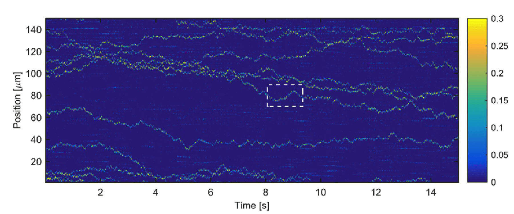
Tracking single, unlabeled, freely diffusing CCMV virions. The color bar indicates the signal level compared to the background. Measurements are taken at 2 KHz, a rate comparable to the timescales of self-assembly.

Publications

Goldfain, A. M. ; Garmann, R. F. ; Jin, Y. ; Lahini, Y. ; Manoharan, V. N. Dynamic Measurements of the Position, Orientation, and DNA Content of Individual Unlabeled Bacteriophages. The Journal of Physical Chemistry B 2016, 120, 6130–6138. Publisher's VersionAbstract

A complete understanding of the cellular pathways involved in viral infections will ultimately require a diverse arsenal of experimental techniques, including methods for tracking individual viruses and their interactions with the host. Here we demonstrate the use of holographic microscopy to track the position, orientation, and DNA content of unlabeled bacteriophages (phages) in solution near a planar, functionalized glass surface. We simultaneously track over 100 individual λ phages at a rate of 100 Hz across a 33 μm × 33 μm portion of the surface. The technique determines the in-plane motion of the phage to nanometer precision, and the height of the phage above the surface to 100 nm precision. Additionally, we track the DNA content of individual phages as they eject their genome following the addition of detergent-solubilized LamB receptor. The technique determines the fraction of DNA remaining in the phage to within 10% of the total 48.5 kilobase pairs. Analysis of the data reveals that under certain conditions, λ phages move along the surface with their heads down and intermittently stick to the surface by their tails, causing them to stand up. Furthermore, we find that in buffer containing high concentrations of both monovalent and divalent salts, λ phages eject their entire DNA in about 7 s. Taken together, these measurements highlight the potential of holographic microscopy to resolve the fast kinetics of the early stages of phage infection.

Faez, S. ; Latin, Y. ; Weidlich, S. ; Garmann, R. F. ; Wondraczek, K. ; Zeisberger, M. ; Schmidt, M. A. ; Orrit, M. ; Manoharan, V. N. Fast, label-free tracking of single viruses and weakly scattering nanoparticles in a nanofluidic optical fiber. ACS Nano 2015, 9 12349-12357. Publisher's VersionAbstract

High-speed tracking of single particles is a gateway to understanding physical, chemical, and biological processes at the nanoscale. It is also a major experimental challenge, particularly for small, nanometer-scale particles. Although methods such as confocal or fluorescence microscopy offer both high spatial resolution and high signal-to-background ratios, the fluorescence emission lifetime limits the measurement speed, while photobleaching and thermal diffusion limit the duration of measurements. Here we present a tracking method based on elastic light scattering that enables long-duration measurements of nanoparticle dynamics at rates of thousands of frames per second. We contain the particles within a single-mode silica fiber having a subwavelength, nanofluidic channel and illuminate them using the fiber’s strongly confined optical mode. The diffusing particles in this cylindrical geometry are continuously illuminated inside the collection focal plane. We show that the method can track unlabeled dielectric particles as small as 20 nm as well as individual cowpea chlorotic mottle virus (CCMV) virions—26 nm in size and 4.6 megadaltons in mass—at rates of over 3 kHz for durations of tens of seconds. Our setup is easily incorporated into common optical microscopes and extends their detection range to nanometer-scale particles and macromolecules. The ease-of-use and performance of this technique support its potential for widespread applications in medical diagnostics and micro total analysis systems.

Garmann, R. F. ; Sportsman, R. ; Beren, C. ; Manoharan, V. N. ; Knobler, C. M. ; Gelbart, W. M. A Simple RNA-DNA Scaffold Templates the Assembly of Monofunctional Virus-Like Particles. JACS 2015, 137, 7584–7587. Publisher's VersionAbstract

Using the components of a particularly well-studied plant virus, cowpea chlorotic mottle virus (CCMV), we demonstrate the synthesis of virus-like particles (VLPs) with one end of the packaged RNA extending out of the capsid and into the surrounding solution. This construct breaks the otherwise perfect symmetry of the capsid and provides a straightforward route for monofunctionalizing VLPs using the principles of DNA nanotechnology. It also allows physical manipulation of the packaged RNA, a previously inaccessible part of the viral architecture. Our synthesis does not involve covalent chemistry of any kind; rather, we trigger capsid assembly on a scaffold of viral RNA that is hybridized at one end to a complementary DNA strand. Interaction of CCMV capsid protein with this RNA-DNA template leads to selective packaging of the RNA portion into a well-formed capsid but leaves the hybridized portion poking out of the capsid through a small hole. We show that the nucleic acid protruding from the capsid is capable of binding free DNA strands and DNA-functionalized colloidal particles. Separately, we show that the RNA-DNA scaffold can be used to nucleate virus formation on a DNA-functionalized surface. We believe this self-assembly strategy can be adapted to viruses other than CCMV.